B/1000/PH
Laboratory microscope for routine and research applications
B/1000/PH - Phase contrast research trinocular microscope
Description
- Laboratory microscope for routine and research applications
- Dye-cast frame, with high stability and ergonomy, for transmitted light observation
- Version for phase contrast analysis.
Transmitted illumination
X-LED8 (8W power)
Description of the general characteristics of the B/1000 series models
B/1000 microscopes, thanks to the long experience achieved in microscopy development, has conceived the new B/1000: a major leap in our technological offer.
As a flag As a flagship instrument, B/1000 originates from customer most demanding feedbacks and needs. Its modularity and versatility will allow to find the perfect place in any clinical or basic reasearch laboratory.
All controls are easily accessible and comfortable also for extended periods of observation.
Highest category of optical equipment among our product range guarantees a sharp and clear view in any situation, while top level mechanical design offers sturdiness and long lifetime.
B/1000 is built on IOS Infinity Corrected optical system, which gives both top-notch optical performances, and the possibility to extend your instrument with the broad range of accessories and modules. X-LED illumination is the best solution to have pure white light, very intense even at higher magnification, and optimum power efficiency given by solid state source. If you search for our best solution to your present and future professional needs, B/1000 is the answer.
Solid Stand – Extra Stability
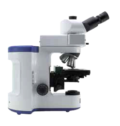
A completely new design and a die-cast aluminum stand offer solidity and durability, even for the most demanding laboratory use. This new microscope can seamlessly be upgraded with many attachments that extend its field of use.
Modularity – Build your own solution
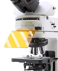
Many worlds in one instrument. Modularity allows to build the desired solution (brightfield, darkfield, phase contrast, material science, fluorescence, motorized automation and so on). B/1000 has the flexibility to help your work the best way.
X-LED White Illumination
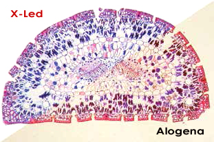
X-LED illumination system is based on a pure white high-efficiency LED and a special optics. It guarantees constant color temperature, no heat, and an extreme electrical consumption efficiency. The whole system is pre-aligned and boasts a lifetime of 50.000 hours.
Light under control

Intelligent control of the microscope illumination: the “AUTO-OFF” function automatically switches the light off after a user-selectable time period. “BOOST” gives an extra high level of illumination for light-demanding applications. “AUTO” allows to store an illumination level, and to maintain it throughout the inspection.
Ergonomia
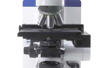
Low position focus and stage controls allow a fast and comfortable operation. Frequently used controls as light intensity adjustment and diaphragm are also placed in the lower part of the stand and enable operation without having to take the eyes off the specimen. All optical heads are equipped with high-point eyepieces and dioptric adjustment, for the best viewing experience.
Comfortable Stage
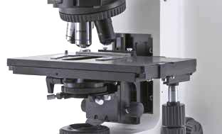
Refined belt-driven stage, with a wide working surface and a highly precise XY movement.
High Quality IOS Optical System
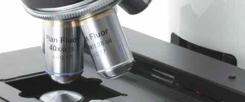
Infinity corrected optical system, based on planachromatic, fluorite, and semiapochromatic objectives, designed to give sharp and clear images, both for the user and the digital camera. Quintuple and sextuple nosepieces give the flexibility to build the optics set that best suits your needs. The system is complete with wide field, high-point eyepieces, with a field number of 24mm.
Ready for Digital Imaging
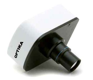
Range of adapters can accommodate for C-mount digital cameras, as well as reflex cameras. Focus adjustment gives perfectly clear digital images. Our cameras include specific software for capturing, measuring, marking and storing your pictures. Optika Vision Pro software allows to perform image acquisition, post-processing, measurements and storage of your images. User can save a preset for later work, or even create a multi-focus composition.
Remote-controlled microscope
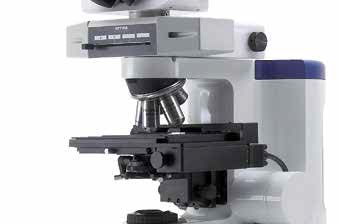
The stage can be remote-controlled through a dedicated software: X, Y, Z axes, as well as nosepiece, can be moved with a single click. Communication protocol is available for interfacing with custom software, such as automated analysis or autofocus.
X-LED benefits
Powerful pure white LED illumination, ideal for brightfield, darkfield and phase contrast applications. Color temperature constant through all the intensity levels. No heat generation, that could damage the specimen.
Factory pre-centering assures uniform illumination over the field of view, yet providing perfect Koehler alignment. Very long lifetime and high power efficiency.
Application fields
 |
Pathology / Cytology
Since B-800 / B-1000 use white LED illumination, they can maintain the same color temperature even if the brightness is changed. “AUTO” function automatically adjusts the light intensity when the objective is changed or the aperture diaphragm is set to a different value. These feautures, along with motorized stage and ergonomic controls, make your workflow easier.
|
 |
Phase Contrast Microscopy
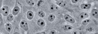
The bright LED illuminator brings a comfortable view in phase contrast with all magnifications. Universal wheel condenser allows to quickly switch between brightfield, darkfield and phase contrast. Ideal for clinical laboratories or fibers (e.g. asbestos) analysis.
|
 |
Fluorescence Microscopy
A new attachment for epi-fluorescence provides the
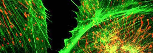
ultimate solution in the field of fluorescence diagnostic. Vibration-free six positions filter wheel with shutter, field and aperture diaphragms, it offers all you need for a complete analysis. Custom filtersets are available and mounted on request. For application where efficiency, rapidity and ease of use are crucial, this model offers also
a LED epi-fluorescence attachment, with very high power standard illuminators.
|
 |
Darkfield Microscopy
Ideal for observing blood cells, diatoms, small insects, bone, fibers, unstained bacteria, yeast, protozoa, mineral and chemical crystals, colloidal particles, dust-count specimens, and thin sections of polymers and ceramics.
|
 |
Material Science
A new attachment designed specifically for metallographic inspection, with dedicated objectives set, for the most complete epi-illumination analysis:
brightfield, darkfield and polarizing view.
|
 |
Polarizing Microscopy
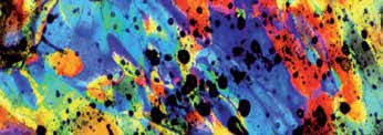
Polarized light microscopy is used in geological applications or also for both natural and industrial minerals, composites such as concretes, ceramics, mineral fibers and polymers, and crystalline or
biological molecules such as DNA, starch, wood and urea. Attachments for a full polarization analysis are available (both for transmitted and incident light), so
it’s possible to look at color fringes right away.
|